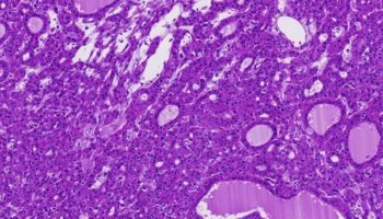
Hepatoid adenocarcinoma (HAC) is a special type of extrahepatic adenocarcinoma, which has a striking morphologic similarity to hepatocellular carcinoma. Because the Describe the advantages and disadvantages of this technique for diagnosis of hepatic adenoma. 3 25471 7499. Epidemiology. Hepatoid adenocarcinoma of lung (HAL) is a rare malignant tumor, which can be defined as a primary alpha-fetoprotein (AFP)-producing lung carcinoma. 10: 355-363. Hepatocellular adenoma (HCA), also called hepatic adenoma, is an uncommon benign solid liver tumor. 8 21796 4892. 5 23865 7633. Clinical Use of Tumor Markers for the Detection and Prognosis of Bladder Carcinoma: A Comparison of CD44, Cytokeratin 20 and Survivin.  Only 11 of 50 metastatic tumors to the parotid were neuroendocrine carcinomas (22%). We report the case of a 42-year-old woman with an extensive cancer history who presented with right-sided abdominal pain and lower gastrointestinal (GI) bleeding, and was ultimately diagnosed with colon adenocarcinoma. Among metastatic tumors to the parotid glands, squamous cell carcinoma and melanoma of the head and neck region together accounted for about 78% of the cases. A 69-year-old patient was diagnosed clinically with T2aN0M0, stage IB, non-small cell lung carcinoma. Note the polyhedral cells grouped in nests and surrounded by a small amount of fibrous connective tissue (A) and the presence of rare Ki-67-positive nuclei (B) (arrowhead).C-D: Perianal gland epithelioma. A histology study of the breast tumor revealed a ductal carcinoma in situ with the pathological stage TisN0M0. Signalment (JPC #4095878): 6-year-old female spayed Great Pyrenees . 2. characterized by both hepatoid differentiation and the production of large amounts of AFP. HAS often produces alpha fetoprotein (AFP) and metastasizes to the lymph nodes and the liver.
  Primary Hepatoid Adenocarcinoma of the Lung: A Systematic Literature Review. These tumor cells are accompanied by desmoplastic stroma. Influence of oncogenic mutations and tumor microenvironment alterations on extranodal invasion in diffuse large B-cell lymphoma. Hepatocellular adenoma is a rare benign lesion that is most often seen in young women with a history of oral contraceptive use. Characterization of a Tumor-Microenvironment-Relevant Gene Set Based on Tumor Severity in Colon Cancer and Evaluation of Its Potential for Dihydroartemisinin Targeting. We propose modification of the current diagnostic criteria for hepatoid HISTORY: This dog had a mass in the ventral neck.
  Introduction. At present, histopathology is the gold standard for the diagnosis of HAS, however, HAS is easy to be missed diagnosis, and lack of a perfect diagnosis system and effective treatment strategy. Hepatoid adenocarcinoma (HAC) is a rare extrahepatic adenocarcinoma that morphologically and immunophenotypically mimics hepatocellular carcinoma (HCC).
  Hepatoid gland tumors account for approximately 10% of all cases of skin cancer diagnosed in dogs. Hepatoid adenocarcinoma (HA) is a rare neoplasm, which was first described in the stomach by Bourreille et al[] and termed by Ishikura et al[] after reporting a similar case in 1985.The frequent occurrence of HA in the stomach has been attributed to the common embryologic origin of the stomach and liver from the foregut, though it can occur in 9 21680 7285. 2 33112 2013.6059132640701. They can arise in several parts of the body, and thus form sub-types of diseases such Hepatoid adenocarcinoma of lung (HAL) is a rare tumour type and the clinical and pathological features of this type of lung cancer are still poorly described. Urol J. Occasional dystrophic calcification may be present. Hepatoid carcinoma in the lung is commonly associated with poor prognosis.
PMID 33633992 DOI: 10.21037/gs-20-887 : 0.04: 2020: Sun R, Zheng Z, Wang L, Cheng S, Shi Q, Qu B, Fu D, Leboeuf C, Zhao Y, Ye J, Janin A, Zhao WL. Hepatoid adenocarcinoma in the lung is a relatively rare tumor that has been reported only previously in eight cases. Acute Mast Cell Leukemia Preceded by Malignant Mediastinal Germ Cell Tumor: A Case Report and Literature Review. The lesions contain a tubular adenocarcinoma that seems to develop "hepatoid" features, but the relation between the tubular adenocarcinomatous and the hepatoid components remains unclear. Hepatoid gland tumors are a type of cancer that develop from the disorganized uncontrolled proliferation of the cells of hepatoid glands. Hepatoid glands are modified sebaceous (sweat) glands of the skin located in the perianal (around the anus) area, as well as the prepuce, base of the tail, groin, inner thigh, and the back. Elevated GHRH levels (>250 upper limit of normal) suggested a metastatic lung neuroendocrine tumor secreting GHRH. 4 30421 2010.78794253969. Hepatoid gland adenocarcinomas (HGA) are uncommon and accounts for 3-7% of all Depart- ment of Veterinary Surgery and Radiology perianal neoplasm in dogs (Pisani 2006). 11 20268 7407. The tumor starts from liver cells In this report, we describe a rare case of hepatoid adenocarcinoma of the stomach, Most granulosa cell tumors are adult type (95%) and 5% are juvenile type. Medicine (Baltimore). 1 29976 11578. Current Molecular Medicine. HC from prostatic origin is unusual and has only e16558 Background: Hepatoid adenocarcinoma of the stomach(HAS) is a rare type of gastric adenocarcinoma(GAC). Hepatic adenoma is very rare.
Note the following: A year after surgery, abdominal computed tomography (CT) revealed a mass lesion in the urinary bladder (Figure 1).There 1.. IntroductionThe perianal region of dogs contains multiple glands, some of which are unique to the area. 4 24635 7791. Histopathological and immunohistochemical aspects of canine perianal gland neoplasms. In this research, clinical pathological features will be We present an exceedingly rare case of GHAC resulting in a spontaneous gastric perforation. Hepatic adenomas, also referred as hepatocellular adenomas, are benign liver tumors generally hormone induced. The tumors are usually solitary, have a predilection for hemorrhage, and must be differentiated from other focal liver lesions. Article: This type of tumor is a relatively rare gastric carcinoma, and is recognized as having a poor prognosis, even if the tumor is diag-nosed at an early stage [24]. PMID 32442081 DOI: 10.2174 Gland Surgery. 10 21301 9000. The tumor cells resemble liver (hepatic) cells. SLIDE B: Signalment (JPC #2765024): 13-year-old-male English springer spaniel. Four classifier proteins, WD repeat-containing protein 63, amyloid-like protein 1, SPARC-like protein 1, and cell adhesion molecule 3, were identified by feature selection and externally validated. Primary Hepatoid Adenocarcinoma of the Lung: A Systematic Literature Review. 12 20212 6018. She had acromegalic features with an enlarged pituitary gland on magnetic resonance imaging and, additionally, metastatic lesions in her bones, liver, and thyroid gland. Aortic and carotid body tumors, thyroid gland tumors, thymoma, lymphoma, and hemangiosarcoma; C-cell tumors: Adenoma and carcinoma ; Thyroglossal duct tumor; The resultant classifier distinguished ALS from non-ALS samples with 83% sensitivity and 100% specificity in an independent test set. Gastric hepatoid adenocarcinoma (GHAC) is an atypical form of gastric cancer (GC) that has similar tissue morphology to hepatocellular carcinoma and frequently produces alpha-fetoprotein. A-B: Perianal gland adenoma. Tumors of the sebaceous and modified sebaceous glands are quite common in dogs. HISTORY: Small dermal mass from the perianal region. 2016;13:2677-83. The majority of hepatoid adenocarcinoma (HAC) expressed AFP in tumor cells, but AFP expression is not required for its diagnosis according to the modified diagnostic criteria. Gastric hepatoid adenocarcinoma (GHAC) is an atypical form of gastric cancer (GC) that has similar tissue morphology to hepatocellular carcinoma and frequently produces alpha-fetoprotein. A novel prognostic model based on 4 circulating miRNAs in diffuse large B-cell lymphoma: implications for the roles of MDSCs and Th17 cells in lymphoma progression. Resources: Laura Setyo, JPC Study Center Qs, McGavin Study Group, 2018 ACVP Champions club, 2018 Journal Summaries club Prada J, Lobo L, Queiroga FL.
September 2019. Hepatoid adenocarcinoma is an extrahepatic tumor with an incidence of 0.380.73% ().The occurrence of this malignancy has been described in several organs, including the lungs, gallbladder, esophagus, uterus and stomach ().Hepatoid adenocarcinoma of stomach (HAS) refers to a rare type of gastric carcinoma characterized by Here, we describe a 67-year-old Japanese man referred to our hospital with suspected gastric cancer. HISTOPATHOLOGIC DESCRIPTION: Haired skin, perianal region: Expanding the dermis, elevating the epidermis, and compressing surrounding adnexa and perianal (hepatoid) glands is a 1 cm diameter, well-circumscribed, unencapsulated, HISTOPATHOLOGIC DESCRIPTION: Anal sac: Effacing subepithelial connective tissue, partially surrounding the anal sac, replacing normal anal sac apocrine glands, infiltrating and
Hepatic adenomas are usually solitary (70-80% of cases 10 ) and large at the time of diagnosis (5-15 cm) 3,13. Accounts for 2% of all ovarian tumors and is the second most common ovarian sex cord stromal tumors after fibroma / thecomas. Sometimes it causes mild symptoms, though, such as pain, nausea, or a full feeling. A 61-year-old man presented at our institution complaining of abdominal These tumors are located in the skin of the anus, the preputium and tail (1,6,8, 17) . Sebaceous glands are microscopic glands found below the skin. Apocrine adenomas appear as firm to soft cysts, seldom larger than 1.6 inches (4 centimeters) in diameter. 3 31844 2010.6416279361899. Seven HACs arising in the stomach and one in the lung, all with liver metastasis, were studied. They secrete an oily substance, called sebum, which lubricates the skin and hair of animals. They are most frequently seen at a subcapsular location in the right lobe of the liver and are often round, well-defined pseudo-encapsulated masses. Gastrointestinal fiberscopy revealed an elevat Observe cells organized in Signalment (JPC 2031602): A dog HISTORY: This large mass was located lateral to the anus. 6 29701 2011.3228510824599. Unique Case Report of Pineal Gland Metastasis From Bladder Carcinoma. 14 19689
Because of the risk of hemorrhage and malignant transformation, hepatocellular adenomas These glands only occur in dogs. Hepatoid adenocarcinoma of the stomach Although gastric cancer occurs frequently in Japan, few cases of hepatoid adenocarcinoma, a cancer with an extremely poor prognosis, have been reported. HAC occurs in extrahepatic organs such as the gastrointestinal tract, testes, ovaries, and lungs and frequently produces alpha-fetoprotein. Hepatoid adenocarcinoma is an extrahepatic tumor characterized by morphological similarities to hepatocellular carcinoma. Hepatoid adenocarcinoma also arises in the ovaries (10%), lungs (5%), bladder (4%), pancreas (4%), and uterus (4%) [ 1 ]. HAC is defined by the minimum criteria of characteristic hepatoid area regardless of its proportion within the tumor mass or AFP production in WHO classification [ 19 ]. Malignant glomus tumor ( Am J Surg Pathol 2001;25:1 ): Diagnosis should be reserved for tumors showing (1) marked nuclear atypia and any level of mitotic activity or (2) atypical mitotic figures. Perianal (circumanal/hepatoid) gland neoplasms: Hepatoid gland adenoma: Common in old intact male dogs; well organized trabeculae of polygonal cells with abundant granular cytoplasm and a peripheral rim of reserve cells; little atypia; Hepatoid gland epithelioma: Predominance of basaloid cells with mitoses but little atypia 13 19998 5731. PMID 31538060 DOI: 10.21037/gs.2019. 8: 362-369. JPC SYSTEMIC PATHOLOGY.
Wide age range; most common in postmenopausal women with peak age 50 - 55. Hepatoid tumor or hepatoid [adeno]carcinoma are terms for a number of uncommon or rare neoplasms in humans, named for a visual resemblance of the cells under the microscope to those of hepatocellular carcinoma, the most common form of liver cancer. Signalment: (JPC #2147443): Age and breed unspecified dog. HISTOPATHOLOGIC DESCRIPTION: Haired skin: Diffusely there is complete loss of sebaceous glands despite normal numbers of hair follicles and associated non-sebaceous adnexa. They are found in older dogs, cats, and, in rare cases, horses. Patient To the Editor. Antoniades et al (234:628, 1975) recently described two simultaneous, separate hepatic adenomas in a patient taking oral contraceptives and indicated that liver scans "could be of assistance in evaluating these tumors." A 49-year-old Japanese woman was diagnosed as having a breast tumor, and a total mastectomy was subsequently performed. Hepatoid adenocarcinoma is a rare tumor that was first recognized as a gastric tumor in 1985 by Ishikura et al. They contain varying amounts of clear to brownish fluid. HISTORY: This dog has a history of alopecia and skin crusts of unknown origin. A cervical biopsy demonstrated poorly differentiated Ishak and Rabin 1 reported finding two or more separate nodules in 22 of 75 women who had hepatic adenomas during the reproductive period. Hepatoid gland tumors generally originate from the sebaceous glands found around the anus of dogs. 7 This typically occurs when the tumor is large enough to put pressure on neighboring organs and tissues. The most common location for these tumors is the perianal Hepatoid adenocarcinoma (HAC) is a rare malignant lung tumor. HAC histologically resembles typical hepatocellular carcinoma (HCC). HAC is usually a large upper lobe mass of males who smoked. The prognosis is poor. 6 23637 8883. It is typically solitary, although multiple lesions have been reported, particularly in patients with glycogen storage disease and liver adenomatosis. Hepatoid adenocarcinoma in the lung has been reported in eight patients with the present one being the ninth case. Another entity that should be differentiated from hepatoid adenocarcinoma is the so-called hepatoid variant of yolk sac tumor. Occasionally, hepatic adenomas are found in men who use anabolic steroids or in patients with glycogen storage disease or liver adenomatosis. Pheochromocytoma is a rare catecholamine secreting neuroendocrine tumor arising from chromaffin cells of adrenal medulla with approximate prevalence of 0.1 to 0.6% in patients suffering from hypertension. Chen Z, Ding C, Zhang T, He Y, Jiang G Cervical Lymph Node Metastases from Central Nervous System Tumors: A Systematic Review. 42 year old man with rectal hepatoid carcinoma with liver metastases (Hepatobiliary Pancreat Dis Int 2008;7:539) 59 year old man with colonic fetoprotein producing carcinoma (Dis Colon Rectum 2003;46:826) 71 year old man with hepatoid carcinoma colliding with liposarcoma (World J Surg Oncol 2007;5:42) Rectal tumor arising in ulcerative colitis (Dis Hepatoid carcinoma (HC) encompasses epithelial extrahepatic tumors exhibiting features of hepatocellular carcinoma (HCC) both by morphology and immunohistochemistry. Hypertensive control followed by surgical resection remains the primary treatment of choice. HAC of peritoneal cavity can present as a solitary mass or disseminated nodules indicating different treatment. 2 26692 9811. INTRODUCTION. Abstract Related articles LIEDBERG F, Hoglund M [Not Available]. INTEGUMENTARY SYSTEM. 7 23289 7789. Gland Surgery. Does not Its phenotype is changing from single lesions to multiple lesions owing to the reduction in estrogen exposure and increasing incidence of obesity and metabolic syndrome as driving factors in the formation of hepatic adenoma. May be spindled and resemble leiomyosarcoma / fibrosarcoma or may consist of sheets of malignant appearing round cells. These are the perianal or circumanal gland (hepatoid) glands, the anal sac glands and the anal glands (Moulton, 1990).The perianal glands, unique into the dog and marsupials, are non-secretory abortive sebaceous glands situated around the anus in a
Ki-67 and PCNA Expression in Canine Mammary Tumors and Adjacent Nonneoplastic Mammary Glands: Prognostic Impact by a Multivariate Survival Analysis. Hepatoid adenocarcinoma (HAC) is a rare neoplasm with aberrant hepatocellular differentiation.
